Bilateral Inverted Papilloma of the Frontal Sinus

INTRODUCTION
Inverted papilloma is a benign tumor of the sinonasal cavity arising from Schneiderian cells. They are known for their high risk of recurrence and potential for malignant degeneration. They typically affect men and occur in the 4th to 6th decades of life, and the most common presenting symptoms is nasal obstruction (1,9). While they typically originate from the lateral nasal wall, they may also originate from the paranasal sinuses with the maxillary and ethmoid sinuses being most commonly involved. The frontal and sphenoid sinuses are less commonly affected with the frontal sinus involved in 1-16% of all cases (1,7).
Staging of inverted papilloma is as follows: Stage I disease is limited to the nasal cavity; Stage II is limited to the ethmoid sinuses and the medial and superior aspects of the maxillary sinus; Stage III involves the lateral and/or inferior aspects of the the maxillary sinuses or extensions to the frontal and/or sphenoid sinuses; Stage IV involves spread outside the nose or sinuses and includes any evidence of malignancy (10).
METHODS: Case Report
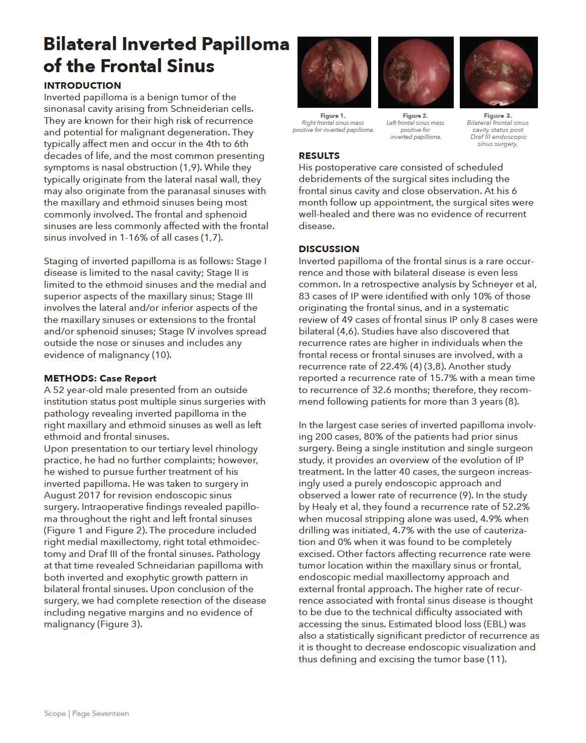
A 52 year-old male presented from an outside institution status post multiple sinus surgeries with pathology revealing inverted papilloma in the right maxillary and ethmoid sinuses as well as left ethmoid and frontal sinuses. Upon presentation to our tertiary level rhinology practice, he had no further complaints; however, he wished to pursue further treatment of his inverted papilloma. He was taken to surgery in August 2017 for revision endoscopic sinus surgery. Intraoperative findings revealed papilloma throughout the right and left frontal sinuses (Figure 1 and Figure 2). The procedure included right medial maxillectomy, right total ethmoidectomy and Draf III of the frontal sinuses. Pathology at that time revealed Schneidarian papilloma with both inverted and exophytic growth pattern in bilateral frontal sinuses. Upon conclusion of the surgery, we had complete resection of the disease including negative margins and no evidence of malignancy (Figure 3).
RESULTS
His postoperative care consisted of scheduled debridements of the surgical sites including the frontal sinus cavity and close observation. At his 6 month follow up appointment, the surgical sites were well-healed and there was no evidence of recurrent disease.
DISCUSSION
Inverted papilloma of the frontal sinus is a rare occurrence and those with bilateral disease is even less common. In a retrospective analysis by Schneyer et al, 83 cases of IP were identified with only 10% of those originating the frontal sinus, and in a systematic review of 49 cases of frontal sinus IP only 8 cases were bilateral (4,6). Studies have also discovered that recurrence rates are higher in individuals when the frontal recess or frontal sinuses are involved, with a recurrence rate of 22.4% (4) (3,8). Another study reported a recurrence rate of 15.7% with a mean time to recurrence of 32.6 months; therefore, they recommend following patients for more than 3 years (8).
In the largest case series of inverted papilloma involving 200 cases, 80% of the patients had prior sinus surgery. Being a single institution and single surgeon study, it provides an overview of the evolution of IP treatment. In the latter 40 cases, the surgeon increasingly used a purely endoscopic approach and observed a lower rate of recurrence (9). In the study by Healy et al, they found a recurrence rate of 52.2% when mucosal stripping alone was used, 4.9% when drilling was initiated, 4.7% with the use of cauterization and 0% when it was found to be completely excised. Other factors affecting recurrence rate were tumor location within the maxillary sinus or frontal, endoscopic medial maxillectomy approach and external frontal approach. The higher rate of recurrence associated with frontal sinus disease is thought to be due to the technical difficulty associated with accessing the sinus. Estimated blood loss (EBL) was also a statistically significant predictor of recurrence as it is thought to decrease endoscopic visualization and thus defining and excising the tumor base (11).
Due to the difficulty of visualization and limitations in surgical access to the frontal sinus, there is currently no universal approach; however, more aggressive surgery has been recommended for bilateral disease (4). Kamel et al looked at treatment of IP in the frontal sinus specifically. Of 119 cases of IP, only 6 involved the frontal sinus with the majority originating from other sinuses and extending into the frontal sinus. They recommend a widened frontal ostium with removal of mucosa +/- drilling of bony attachment site (2). In a retrospective review of 9 cases of frontal sinus IP, those with lateral wall of frontal recess involvement underwent endoscopic Draf II A frontal sinusotomy while those with either lateral and posterior wall or medial and posterior wall of the frontal recess involvement plus frontal infundibulum involvement underwent a Draf II B surgery. Draf III surgery was utilized in 2 cases of frontal IP in which the posterior wall of the frontal recess and frontal infundibulum or medial, lateral and posterior walls of the frontal recess and frontal infundibulum were involved (5).
While surgery is currently the gold standard of treatment, possible future treatment may also include topical agents which are currently being investigated. In study of 8 patients with IP involving the frontal sinus, all received an application of 5-FU postoperatively. Their results showed no evidence of recurrence after 42 months in all cases (12). While this data is one of the first of its kind, it leaves open the door to further research in this treatment modality.
CONCLUSSION
Bilateral frontal sinus disease is an extremely rare presentation for inverted papilloma. Due to the potential for recurrence and malignant transformation of IP, it is important to be diligent to perform complete resection of disease with negative margins. Surgical resection using endoscopic surgery has been shown to provide consistent results with low rates of recurrence.
References:
Vorasubin N, Vira D, Suh JD, Bhuta S, Wang MB. Schneiderian papillomas: Comparative review of exophytic, oncocytic, and inverted types. American Journal of Rhinology & Allergy. 2013;27(4):287-292. Kamal RH, Abdel Fattah AF, Awad AG. Origin oriented management of inverted papilloma of the frontal sinus. Rhinology. 2012;50(3):262-8. Wu X, Sun D, Meng X, Yuan Y. The management of sinonasal inverted papilloma by endoscopic surgery: an analysis of 54 cases. Lin chung Er Bi Yan Hou Tou Jing Wai Ke Za Zhi.2014;28(22):1783-1788. Walgama E, Ahn C, Batra PS. Surgical management of frontal sinus inverted papilloma: a systematic review. Laryngoscope. 2012; 122(6):1205-1209. Zhang L, Han DM, Wang CS, et al. Endoscopic management of the inverted papilloma involving frontal sinus and its drainage pathway. Zhonghua Er Bi Yan Hou Tou Jing Wai Ke Za Zhi. 2008;43(1):22-26. Schneyer MS, Milam BM, Payne SC. Sites of attachment of Schniederian papilloma: a retrospective analysis. Int Forum Allergy Rhinol. 2011;1(4):324-328. Shohet JA, Duncavage JA. Management of the frontal sinus with inverted papilloma. Otolaryngol Head Neck Surg. 1996;114:649–652. Kim DY, Hong SL, Lee CH, et al. Inverted papilloma of the nasal cavity and paranasal sinuses: a Korean multicenter study. Laryngoscope. 2012;122(3):487-494. Lawson W, Patel ZM. The evolution of management for inverted papilloma: an analysis of 200 cases. Otolaryngol Head Neck Surg. 2009;140(3):330-335. Krouse JH. Development of a staging system for inverted papilloma. Laryngoscope. 2000 Jun;110(6):965-8. Healy, D. Y. J., Chhabra, N., Metson, R., Holbrook, E. H., & Gray, S. T. Surgical risk factors for recurrence of inverted papilloma. Laryngoscope. 2016;126(4):796–801. Adriaensen GF, van der Hout MW, Reinartz SM, et al. Endoscopic treatment of inverted papilloma attached in the frontal sinus/recess. Rhinology. 2015;53(4):317-324.
Emily Johnson , DO AOCOO-HNS Otolaryngologist/ENT
